CCLF_NEURO_0010_T BioVector® 人类原代胶质母细胞瘤/神经肿瘤患者来源细胞系Human Patient-Derived Glioblastoma/Neuro Oncology Cell Line
- 价 格:¥99860
- 货 号:BioVector® CCLF_NEURO_0010_T
- 产 地:北京
- BioVector NTCC典型培养物保藏中心
- 联系人:Dr.Xu, Biovector NTCC Inc.
电话:400-800-2947 工作微信:1843439339 (QQ同号)
邮件:Biovector@163.com
手机:18901268599
地址:北京
- 已注册
BioVector® CCLF_NEURO_0010_T 人类原代胶质母细胞瘤/神经肿瘤患者来源细胞系 / BioVector® CCLF_NEURO_0010_T Human Patient-Derived Glioblastoma/Neuro Oncology Cell Line
背景与来源说明 / Context & Provenance: BioVector® CCLF_NEURO_0010_T 是由 Broad 研究所(Broad Institute)癌症细胞系工厂(Cancer Cell Line Factory, CCLF)项目直接建立的珍稀人源患者来源细胞模型(Patient-Derived Cell Line, PDCL)。该细胞系直接分离自人类恶性原发性中枢神经系统肿瘤——**胶质母细胞瘤(Glioblastoma, GBM)**患者的手术切除活检标本。作为 Broad 研究所 CCLE(癌症细胞系百科全书)高保真临床转化医学研究计划的一部分,该细胞系现作为核心活体生物样本保藏于全球核心临床基因组联合库。本技术档案严格基于 Broad 研究所分子病理数据库、CCLE 测序图谱以及患者来源异种移植(PDX)质控参数进行双语编制。
通用定义BioVector® CCLF_NEURO_0010_T 是一种源自人类原发恶性脑肿瘤的患者来源胶质母细胞瘤干细胞样(GSC)多维生长型模型。为了完美复刻临床患者体内真实的胶质母细胞瘤空间异质性,该细胞系在体外完全杜绝了人工富血清培养基的干扰,采用支持未分化神经前体细胞增殖的无血清、富含生长因子的特制微环境体系,表现为特征性的贴壁兼具悬浮神经球(Neurospheres)生长模式。
在现代转化医学、精准医疗设计以及针对高级别胶质瘤(HGG)多药耐药机制的研究中,CCLF_NEURO_0010_T 具备极高的转化医学预测价值。由于其保留了原发病灶最初的复杂表观遗传修饰与基因组异质性,它高度呈现了胶质母细胞瘤的关键分子病理特征(如典型的染色体拷贝数异常、受体酪氨酸激酶通路异常以及特征性的代谢重塑)。这使其成为国际上用于高通量筛选新型小分子靶向阻断剂、开发脑靶向递送纳米载体、以及进行个体化肿瘤免疫新抗原预测的标准临床前保真微沙盘。
BioVector® CCLF_NEURO_0010_T 技术与细胞学特征
1. 细胞形态与空间三维生长特性
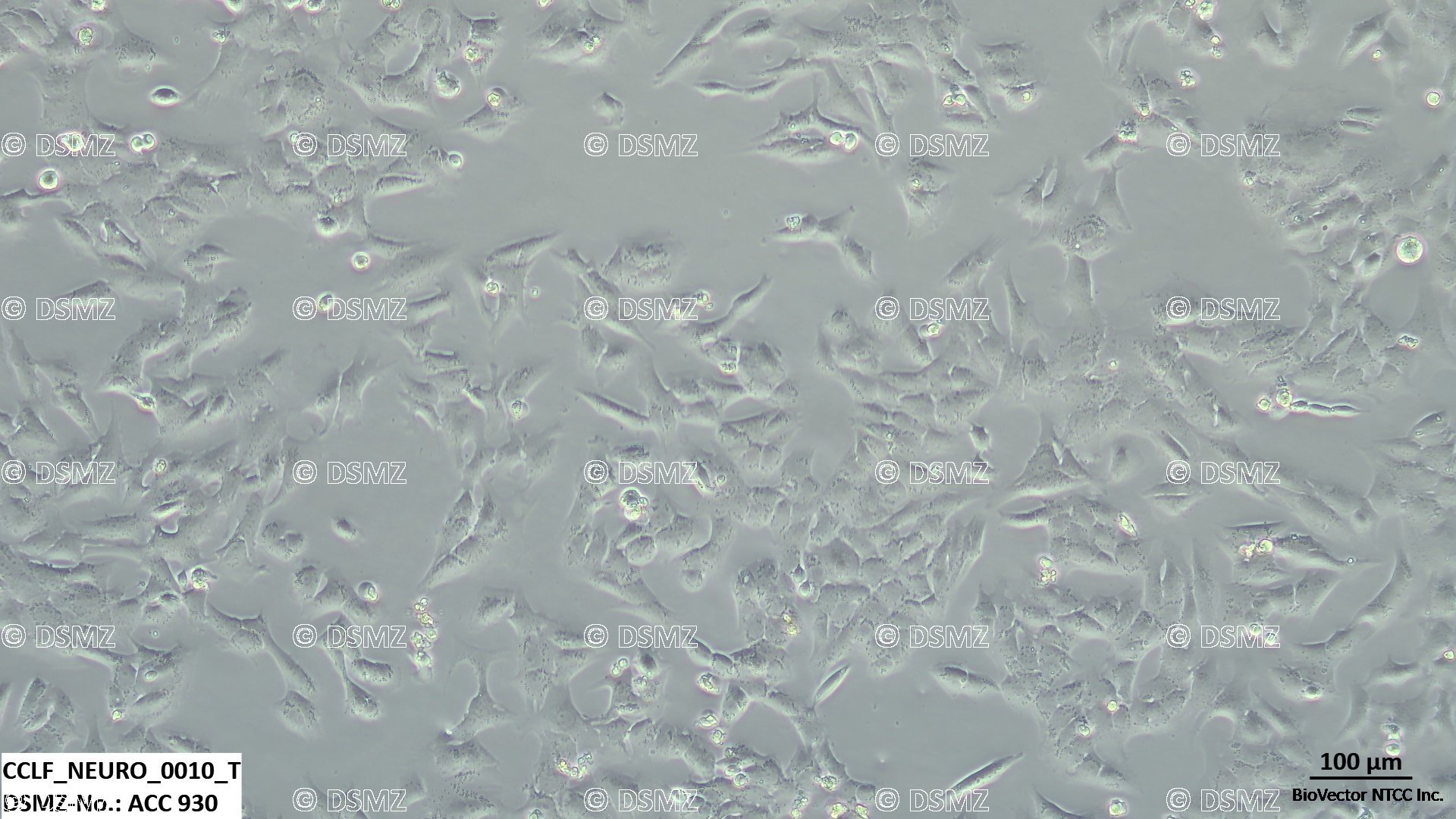
显微形态与聚簇表型 在未包被的超低黏附(Low-attachment)无血清培养容器中,细胞具有极强的自发空间聚集倾向,会逐渐发育成外观立体、边界清晰且高度折光的三维恶性神经肿瘤球结构(Tumor Neurospheres)。若将其接种于包被了基质胶(Matrigel)或聚鸟氨酸/层粘连蛋白(Poly-Ornithine/Laminin)的基质表面时,细胞则会平铺伸展,表现为两极或多极延伸的神经上皮样或星形胶质样前体细胞形态。
干性标记物稳态表达 处于未分化的原始克隆状态。细胞表面及胞质内稳定呈现神经干细胞标志物 Nestin(巢蛋白)、SOX2 以及 CD133(PROM1) 的高丰度强阳性表达,具备在体外长期维持自我更新(Self-renewal)和潜在多向分化的能力。
分子遗传学高保真谱系(核心转化医学资产) 该模型通过全外显子组测序(WES)和 RNA-seq 严格鉴定,其单核苷酸变异(SNV)、染色体拷贝数畸变(CNA)以及特定的驱动基因突变谱与患者原始临床实体肿瘤切片高度吻合,在长期的体外有限传代中展现出极其稳定的病理学基因指纹特征。
2. 严格的神经干细胞专用培养条件与操作指南
为了防止该患者来源细胞系发生不可逆的自发分化,在常规维持与扩增过程中,绝对不可以加入任何成分的胎牛血清(FBS)。日常养护必须严格遵循以下专属配方:
推荐完全无血清培养基配方(神经干细胞专用) * 50% 优质 DMEM/F12 基础培养基 + 50% Neurobasal 基础培养基(或全 Neurobasal 基础培养基)
2 percent BioVector® B27 补剂(不含维生素A,即 B27 Supplement, Minus Vitamin A)
1 percent BioVector® N2 补剂(N2 Supplement)
2 mM GlutaMAX 稳定型谷氨酰胺替换物
绝对必须临用前单独添加的核心重组细胞因子:20 ng/mL 人重组表皮生长因子(Recombinant Human EGF) 以及 20 ng/mL 人重组碱性成纤维细胞生长因子(Recombinant Human bFGF)。
培养环境参数 37 摄氏度,含有 5 percent 二氧化碳(CO2)的恒温恒湿培养箱。
倍增时间与低剪切力消化传代规范 * 传代周期 保留了患者原代原位生长相对隐匿、平缓的特征,倍增时间通常在 48 到 72 小时。当悬浮神经球的平均直径达到 150–200 μm 时(通常每 5 到 7 天)需要及时传代。
消化流程解离 吸出含有细胞球的悬液,1500 rpm 离心 5 分钟捕获沉淀。弃去上清后,加入标准的 Accutase(广谱细胞酶切消化液,严禁使用剧烈粗暴的普通胰蛋白酶)于 37 摄氏度温和浸润 3 到 5 分钟。
轻柔机械打散 使用标准的 1 mL 微量移液器枪头,轻柔吹打 5–8 次,将三维肿瘤球打散为单细胞或 2–3 个细胞的小微团。加入等体积的完全无血清基础培养基终止消化后,按照 1:2 至 1:3 的比例常规分装到新的超低黏附容器内。
主要科研应用
1. 胶质母细胞瘤精准分子靶向与靶点敏感性功能基因组学筛选
保真耐药沙盘建立 BioVector® CCLF_NEURO_0010_T 作为不含任何人工血清和未发生体外过度演化变异的临床高度保真微沙盘,被广泛用于精准测试各种受体酪氨酸激酶(RTK)级联阻断剂、DNA 损伤修复(DDR)抑制剂的杀伤效率。可真实模拟患者临床治疗过程中的多药耐药性(MDR)病理状态。
2. 胶质瘤干细胞 3D 侵袭力与脑组织微环境模拟
3D Matrigel 侵袭分析 利用该细胞自发形成的三维神经球,将其包裹于不同组分的基质胶滴中,通过活细胞影像系统动态追踪肿瘤球边缘细胞向外放射状浸润、迁移的速度,用以评估小分子化合物阻断脑胶质瘤浸润性生长的药效学特征。
技术指标简表
实验操作防坑指南
细胞因子的降解严防:完全培养基中的重组细胞因子(EGF & bFGF)在 37 摄氏度下半衰期极短(通常仅 24–48 小时)。因此,切勿一次性配制过大体积的配方液。强烈建议采取基础液在 4 摄氏度避光封存,每次传代或换液时,仅吸取当次所需的体积,并现场单独加入 fresh 细胞因子,以保证干性维持。
血清污染即死红线:即便是在短暂的传代、洗涤或冻存步骤中,也绝对不可以接触含有胎牛血清(FBS)的液体。一旦细胞意外暴露于血清中,会迅速触发胶质母细胞瘤干细胞向成熟星形胶质细胞不可逆地大面积终末分化,导致整个科研报告基因表达图谱及干性增殖能力瞬间崩盘。
BioVector® CCLF_NEURO_0010_T Human Patient-Derived Glioblastoma/Neuro Oncology Cell Line
General DefinitionBioVector® CCLF_NEURO_0010_T is a highly unique and clinically reflective Patient-Derived Cell Line (PDCL) representing a high-fidelity model of human Glioblastoma (GBM). This line was established directly from patient clinical surgical resections or biopsy tissues by the Cancer Cell Line Factory (CCLF) initiative at the Broad Institute. As an integral component of the Broad Institute's high-fidelity translational oncology frameworks (CCLE project), this lineage is curated within core global genomic reference infrastructure. This technical dataset profile is compiled in accordance with established molecular pathology criteria and patient-derived xenograft (PDX) validation controls.
In translational neuro-oncology, therapeutic screening, and targeted drug resistance profiling, CCLF_NEURO_0010_T functions as an invaluable preclinical tool. Void of artificial serum adaptation, it preserves the intricate epigenetic alterations and cellular heterogeneity found in primary patient high-grade gliomas. It stably reflects the hallmark driving mutations and genomic patterns native to the parent tumor tissue, making it a premier choice for high-throughput screens of small-molecule inhibitors, blood-brain barrier-penetrant discovery, and personalized vaccine-neoantigen discovery pipelines.
BioVector® CCLF_NEURO_0010_T Technical & Cytological Specifications
1. Morphology and Multidimensional Spatial Proliferation
Microscopic Presentation & Heterogeneity Exhibits a versatile growth profile sensitive to its attachment environment. In ultra-low attachment vessels, cells exhibit a powerful natural compulsion to aggregate into highly refractile, three-dimensional malignant tumor neurospheres. When introduced onto surfaces coated with extracellular matrix anchors (such as Matrigel or Poly-L-Ornithine/Laminin), cells readily attach and spread out, presenting as complex bipolar or multipolar neural progenitor configurations with active cytoplasmic extensions.
Stemness Verification Retains an undifferentiated, highly clonogenic phenotype. Both monolayers and floating spheres uniformly display intense intracellular and membrane-bound signals for core neural stem cell biomarkers Nestin, SOX2, and CD133 (PROM1), preserving their capacity for continuous self-renewal.
Patient-Fidelity Genomic Fingerprint (Core Asset) Extensively validated via Whole Exome Sequencing (WES) and RNA-seq. Its single nucleotide variants (SNV) and copy number alterations (CNA) precisely match the parent tumor block, demonstrating exceptional preservation across limited serial passages in vitro.
2. Strict Serum-Free Stemness Cultivation Protocols
To avert the immediate, irreversible terminal differentiation of this patient-derived model, never expose these cultures to Fetal Bovine Serum (FBS) or standard serum-supplemented media. Cultivation must strictly adhere to the following defined serum-free framework:
Formulated Serum-Free Complete Medium (Neural Stem Cell Specific) * 50% premium DMEM/F12 basal medium + 50% Neurobasal medium (or 100% Neurobasal formulation).
2 percent BioVector® B27 Supplement, Minus Vitamin A (essential to suppress unwanted retinoic acid-driven differentiation cascades).
1 percent BioVector® N2 Supplement.
2 mM GlutaMAX stable dipeptide.
Mandatory Extracellular Growth Factors (Must be added fresh): Enriched with 20 ng/mL Recombinant Human EGF and 20 ng/mL Recombinant Human bFGF.
Incubation Parameters 37 degrees Celsius under a humidified environment supplemented with 5 percent Carbon Dioxide (CO2).
Kinetics and Low-Shear Dissociation Routines Reflecting its authentic primary lineage, the model expands at a measured pace, exhibiting an average cell doubling window of 48 to 72 hours.
Neurosphere Dissociation Routine When tumor spheres achieve average diameters of 150–200 μm or show dense central clustering (typically every 5 to 7 days), collect the sphere-laden media and harvest via centrifugation at 1500 rpm for 5 minutes.
Gentle Enzymatic Cleavage Resuspend the pellet in standard Accutase solution (avoid violent, harsh Trypsin preparations) and incubate at 37 degrees Celsius for 3 to 5 minutes. Gently pass through a 1 mL pipette tip 5 to 8 times to break down the 3D structures into single cells or tiny 2–3 cell microscopic matrices. Quench using an equal volume of serum-free complete media, and seed out at a routine 1:2 to 1:3 ratio into ultra-low attachment flasks.
Primary Research Applications
1. Translational Precision Medicine & Innate Chemoresistance Assays
Patient-Fidelity Drug Efficacy Profiling BioVector® CCLF_NEURO_0010_T serves as an authentic in vitro framework devoid of artificial serum stress. It is widely deployed to evaluate the therapeutic thresholds of next-generation RTK inhibitors and DNA damage repair (DDR) targeting agents, bypassing the high false-positive rates typical of over-passaged cell models.
2. 3D Invasion and Matrix Dynamics Assays
3D Matrigel Infiltration Modeling Intact tumor spheres can be embedded directly within extracellular matrix matrices to observe the radial infiltration kinetics of invading cells via live-cell time-lapse photography, establishing an essential assay platform to test anti-metastatic/anti-invasive candidates.
Technical Data Summary
BioVector NTCC质粒载体菌株细胞蛋白抗体基因保藏中心
电话:400-800-2947
工作QQ/微信同号:1843439339
网址http://www.biovector.net
- 公告/新闻