Col-GFP-HSC BioVector® 大鼠胶原蛋白启动子报告基因肝星状细胞系Rat Collagen Promoter Reporter Hepatic Stellate Cell Line
- 价 格:¥99860
- 货 号:BioVector® Col-GFP-HSC
- 产 地:北京
- BioVector NTCC典型培养物保藏中心
- 联系人:Dr.Xu, Biovector NTCC Inc.
电话:400-800-2947 工作微信:1843439339 (QQ同号)
邮件:Biovector@163.com
手机:18901268599
地址:北京
- 已注册
BioVector® Col-GFP-HSC 大鼠胶原蛋白启动子报告基因肝星状细胞系 / BioVector® Col-GFP-HSC Rat Collagen Promoter Reporter Hepatic Stellate Cell Line
背景与来源说明 / Context & Provenance: BioVector® Col-GFP-HSC 是一种专为实时定量监测肝纤维化、细胞活化以及药物筛选而设计的高度工程化功能报告细胞模型。该细胞系基于原代或永生化的大鼠(Rattus norvegicus)肝星状细胞(Hepatic Stellate Cells, HSCs),通过慢病毒或稳定转染技术,将绿色荧光蛋白(GFP)报告基因牢牢置于I型胶原蛋白 链(Collagen Type I , Col1a1)启动子的转录调控之下。本技术档案严格基于细胞外基质(ECM)病理学、纤维化分子筛查标准以及稳定株质控参数进行双语编制。
通用定义BioVector® Col-GFP-HSC 是一种源自大鼠的、带有纤维化活化特异性报告系统的永生化肝星状细胞(HSC)贴壁生长型细胞系。肝星状细胞是介导各种慢性肝损伤引发肝纤维化(Liver Fibrosis)的核心效应细胞。
在正常/静息(Quiescent)状态下,该细胞系保持低水平的基线荧光。然而,当受到促纤维化核心因子(如转化生长因子-β1, TGF-β1)刺激、机械硬度刺激或发生自发性贴壁活化时,细胞会迅速向肌成纤维细胞(Myofibroblast)表型转分化。此时,内源性 Col1a1 启动子被强力激活,直接驱动下游 GFP 的高丰度表达。这种“转录激活与绿色荧光增强”的同步耦联特性,使其无需通过复杂的免疫印迹(WB)或固定染色,即可在体外活细胞状态下通过荧光强度直接、定量地反映 HSC 的活化水平。这使其成为国际上用于高通量筛选抗纤维化小分子药物、评估肝毒性、以及解析星状细胞表型转换表观遗传学机制的黄金报告工具株。
BioVector® Col-GFP-HSC 技术与细胞学特征
1. 细胞形态与动态报告功能特征
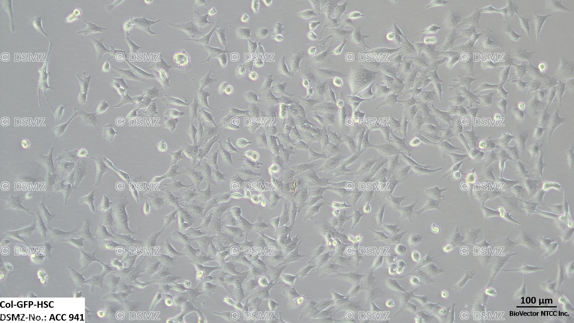
显微形态与表型转换 属于贴壁生长型细胞。在刚复苏或低活化基线状态下,细胞呈现扁平、多角形或带有细长突起的星状形态,胞质内常含有脂滴残留。随着活化程度加深,细胞会转变为宽大的、具有强分泌特征的梭形肌成纤维细胞样形态。
Col-GFP 动态报告系统机制(核心学术亮点)
静息/未活化状态 GFP 荧光信号极弱或呈阴性,代表 Col1a1 启动子处于抑制或低转录活性状态。
活化/诱导状态 在加入 TGF-β1(推荐浓度 2–5 ng/mL)诱导 24–48 小时后,细胞内会爆发性产生明亮的绿色荧光信号(激发的最大波长 ,发射的最大波长 )。荧光强度的对数增长与内源性 I 型胶原蛋白 mRNA 的转录丰度呈严格的正相关关系。
特征性纤维化标记物 随着 GFP 信号的增强,细胞同步高表达 α-平滑肌肌动蛋白(α-SMA)、结蛋白(Desmin)以及纤维粘连蛋白(Fibronectin)。
2. 培养条件与操作指南
推荐基础培养基 高糖 DMEM 基础培养基(含 4.5 g/L 葡萄糖、L-谷氨酰胺及丙酮酸钠,占比 85 到 90 percent)。
推荐血清体系 10 到 15 percent 高质量的 BioVector® Fetal Bovine Serum 胎牛血清。
稳定筛选维持压 为防止长期传代过程中报告基因发生自发性基因沉默或丢失,建立稳定单克隆后,在常规维持培养基中通常需要添加特定浓度的筛选抗生素(通常为 1–2 g/mL 嘌呤霉素 Puromycin 或 200–400 g/mL 选定 G418,视具体工程株构建抗性而定;复苏初期前 24 小时建议不加药)。
培养环境参数 37 摄氏度,含有 5 percent 二氧化碳(CO2)的恒温恒湿培养箱。
倍增时间与传代避坑雷区 细胞增殖相对活跃,倍增时间通常在 24 到 36 小时。
防自发活化控密度法 肝星状细胞具有极强的“接触或硬度依赖性自发活化”倾向。日常维持传代时,绝对不能让细胞 100% 完全长满汇合。当细胞单层密度达到 75 到 80 percent 时必须立刻传代。
常规消化流程 使用 0.25 percent 胰蛋白酶加 0.02 percent EDTA 消化液处理 1–2 分钟。镜下观察到细胞变圆、开始脱离瓶壁时,立即加入含血清的完全培养基终止消化,常规按照 1:3 至 1:5 的比例进行常规分瓶。
主要科研应用
1. 抗肝纤维化(Anti-Fibrotic)小分子药物高通量表型筛选
活细胞荧光药效定量 借助 BioVector® Col-GFP-HSC,科研人员可直接将细胞接种于 96 孔或 384 孔黑壁底透板中。通过预先加入促纤维化因子(如 TGF-β 或 IL-13)建立纤维化沙盘,随后加入药物库。利用活细胞微孔板荧光分析仪直接读取 GFP 的降幅,从而实现非破坏性、高通量的抗纤维化活性药物大规模初筛。
2. 肝脏微环境基质硬度(Matrix Stiffness)力学信号转导研究
力学-转录耦联机制机制 该细胞系常被接种于不同水凝胶硬度表面(如 0.5 kPa 正常肝脏硬度 vs. 32 kPa 晚期肝硬化硬度),用以直接可视化阐明整合素(Integrins)以及 YAP/TAZ 信号通路是如何感知细胞外基质力学变化并直接反向驱动 Col1a1 启动子开放的病理生理进程。
技术指标简表
实验操作防坑指南
铺板底物警戒:如果您希望维持该细胞在较低的基线活化水平(静息态),请尽量避免将其直接长期种植在未经处理的超硬塑料培养皿表面。在可能的情况下,使用包被了鼠尾 I 型胶原凝胶软基质或低黏附表面的器皿,能大幅压低背景自发荧光。
血清批次控制:不合格的胎牛血清中可能含有微量的内源性促纤维化生长因子(如活跃的 TGF-β 变体),会直接推高 GFP 的基线背景。因此,强烈建议使用经 BioVector® 严格质控、内源因子极低的特级胎牛血清进行长期维持。
BioVector® Col-GFP-HSC Rat Collagen Promoter Reporter Hepatic Stellate Cell Line
General DefinitionBioVector® Col-GFP-HSC is an engineered, highly functional rat hepatic stellate cell (HSC) reporter line tailored for real-time monitoring of fibrogenesis, cell activation kinetics, and high-throughput anti-fibrotic drug discovery. This line incorporates a specialized genetic cassette where the transcription of the Green Fluorescent Protein (GFP) reporter molecule is strictly directed by the promoter sequence of the Collagen Type I (Col1a1) gene. This technical catalog dataset is structured based on verified extracellular matrix (ECM) pathology research parameters and stable line quality control matrices.
In chronic hepatic pathophysiology, hepatic stellate cells serve as the primary cellular drivers responsible for the excessive deposition of scar tissue leading to liver fibrosis and cirrhosis. In its quiescent basal state, the BioVector® Col-GFP-HSC line displays a negligible fluorescent signal. However, upon exposure to master fibrotic cytokines (such as TGF-β1) or when triggered by substrate stiffness, the cells undergo transdifferentiation into alpha-smooth muscle actin (α-SMA)-positive myofibroblasts. This phenotypic switch drives the immediate activation of the Col1a1 promoter, prompting a synchronized upsurge in green fluorescence. This precise coupling allows investigators to quantify cellular activation states directly in living cultures without fixative processing, making it a gold-standard indicator for anti-fibrotic therapeutic validation.
BioVector® Col-GFP-HSC Technical & Cytological Specifications
1. Morphological & Dynamic Reporter Characteristics
Phenotypic Transdifferentiation Classified as an adherent cell line. In unactivated or early post-thaw stages, it displays a flat, polygonal, or classically stellate appearance adorned with slender cytoplasmic extensions. Upon myofibroblastic activation, cells stretch into expansive, highly secretory, spindle-shaped configurations.
Col-GFP Mechanistic Kinetics (Academic Benchmark)
Quiescent Baseline Matrix Demonstrates dim or completely negative GFP expressions, indicative of a tightly repressed or low-turnover Col1a1 transcript domain.
Activated Inducible Array Treatment with recombinant TGF-β1 (typically 2–5 ng/mL) for 24–48 hours triggers a robust activation cascade, producing intense green fluorescence (, ). The exponential elevation of fluorescent intensity correlates linearly with endogenous Collagen Type I mRNA copy counts.
Fibrotic Profile Verification GFP expression is tightly correlated with the concurrent upregulation of native fibrotic axis structural proteins, including α-SMA, Desmin, and Fibronectin.
2. Cultivation & Maintenance Guidelines
Recommended Basal Media Formulated in high-glucose DMEM containing 4.5 g/L glucose, L-glutamine, and sodium pyruvate (85 to 90 percent).
Serum Supplementation Augmented with 10 to 15 percent performance-certified BioVector® Fetal Bovine Serum.
Maintenance Antibiotic Selection Pressure To avoid spontaneous epigenetic silencing or transgene dropout across extended serial passage cycles, the complete growth medium should be enriched with selection antibiotics (typically 1–2 g/mL Puromycin or 200–400 g/mL G418, depending on the specific resistance profile of the engineering clone; omit antibiotic selection for the initial 24 hours post-thaw).
Incubation Parameters 37 degrees Celsius under a humidified environment supplemented with 5 percent Carbon Dioxide (CO2).
Kinetics & Seeding Traps Exhibits an active proliferation pace with an average cell doubling window of 24 to 36 hours.
Prevention of Spontaneous Activation Hepatic stellate cells are exceptionally prone to contact-induced or stiffness-mediated spontaneous transdifferentiation. Therefore, never allow the culture monolayer to reach 100 percent saturation confluence. Subculture immediately once densities touch 75 to 80 percent.
Enzymatic Dissociation Routine Treat the monolayer with standard 0.25 percent Trypsin + 0.02 percent EDTA for 1–2 minutes. As cells round up and detach under microscopic check, immediately arrest dissociation using serum-fortified complete medium and split at a routine 1:3 to 1:5 ratio into fresh vessels.
Primary Research Applications
1. High-Throughput Screening (HTS) of Anti-Fibrotic Small Molecule Libraries
Live-Cell Phenotypic Quantification BioVector® Col-GFP-HSC can be seeded directly into 96-well or 384-well black-walled, clear-bottom plates. Following fibrotic priming with target cytokines (e.g., TGF-β), test candidate chemical compounds can be added. Real-time suppression of GFP intensity can be tracked via automated live-cell fluorescent plate readers, facilitating non-destructive high-throughput screens.
2. Mechanotransduction and Matrix Stiffness Crosstalk Assays
Mechanical-to-Transcriptional Coupling Mapping The cells are frequently cultured across customizable polyacrylamide or hydrogel surfaces of variable elastic moduli (e.g., 0.5 kPa mimicking healthy liver vs. 32 kPa modeling cirrhotic tissue) to visualize how integrin clusters and downstream YAP/TAZ mechanosensitive complexes drive chromatin remodeling and open the Col1a1 promoter locus.
Technical Data Summary
BioVector NTCC质粒载体菌株细胞蛋白抗体基因保藏中心
电话:400-800-2947
工作QQ/微信同号:1843439339
网址http://www.biovector.net
- 公告/新闻